Presented by Charles & Linda Raabe
Mactan Island, The Philippines
© 2008 All Rights Reserved
Also see:
The Caridean Shrimp of the Central Visayan Islands
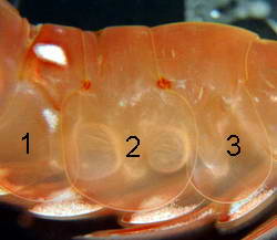 How does one tell the difference between the Caridean (true
shrimp) and the Penaeids (prawns)? The quickest method is to
simply examine the second abdominal segment. If segment two
overlaps both segments one and three, then you have a Caridean
(shrimp). If segment two overlaps only segment three, then you
have a Penaeid (prawn).
How does one tell the difference between the Caridean (true
shrimp) and the Penaeids (prawns)? The quickest method is to
simply examine the second abdominal segment. If segment two
overlaps both segments one and three, then you have a Caridean
(shrimp). If segment two overlaps only segment three, then you
have a Penaeid (prawn).Caridoid facies :
The morphological characteristics that adapt a true shrimp to swimming is known as the Caridoid facies. Carid (latin) means shrimp with the suffix oid meaning "-like" and facies refers to the general appearance. The crustaceans that most fully exhibit the caridoid facies are those species found in the Malacostracan super order Eucarida (Eu, "true", Carrid,"shrimp"). The components of the caridoid facies involve hydrodynamic adaptations that make shrimp highly streamlined and suited for swimming. The paddle like pleopods power forward swimming and in some caridoids, oar like outer branches of the thoracid legs (exopods) also aide in forward swimming. The backward tail flipping (retrograde escape response) of a frightened or startled shrimp is another form of swimming to which several caridoid features are involved. Propulsion is provided by rapid flexing of the abdomen which pushes forward the tail fan uropods propelling the shrimp swiftly backwards.
In true shrimp, a cuticular cover, the carapace, extends over and covers the thoracid segments that streamlines the shrimp. The dorsal midline of the cephalothorax is often produced into a dorsal carina (keel) that looks and acts much like the keel of a ship, but in shrimp, the "keel" is upside down yet serves the same function by providing stability as the shrimp moves through the water. An extension of the dorsal carina is the rostrum, a sword like blade that presumably functions as another aide in stability. The flat antennal scales (exopods of the second antennae) spread and tilt during swimming, acting as both rudders and brakes. The uropods of the tail fan are also important steering devices during backward propulsion.
Stalked eyes and biramous antennule (first antenna) are adaptations of mobile shrimp. The compound eye located on a movable stalk can detect movement and form images over a wide field of vision. The area that they can cover with such stalked eyes became quite apparent during my efforts to catch live specimens as I quickly became aware of the fact that there is no such thing as sneaking up behind a shrimp. The outer branch of the antennule caaries the aesthetascs which are sensitive to dissolved compounds in the water, a sense of "smell" if you will. The long antennae carries a variety of tactile or touch receptors as well as chemoreceptors sensitive to higher concentrations of chemical stimuli able to sweep all around the shrimp in order to detect and recognize nearby objects. This ability to reach out and touch can be observed with captive shrimp that have grown accustomed to hand feeding. But before approaching the hand that literally feeds them, they will first sweep their antennae over your hand or fingers as their means to taste and touch test before approaching closer. This ability also ensures their safety in the wild as it gives them a means to detect non-moving hazards that their motion sensitive eyes can not detect.
The caridoid form is considered to be a very successful one within the marine habitats but in terms of numbers of species, the carideans are by far the most successful. Such high biological diversity reflects their much wider range of habitats in comparison with other caridoid groups. There can be little doubt that the caridoid body plan, having been modified in a variety of ways, has been the basis from which extensive invasion and diversification in the benthic environment has occurred.
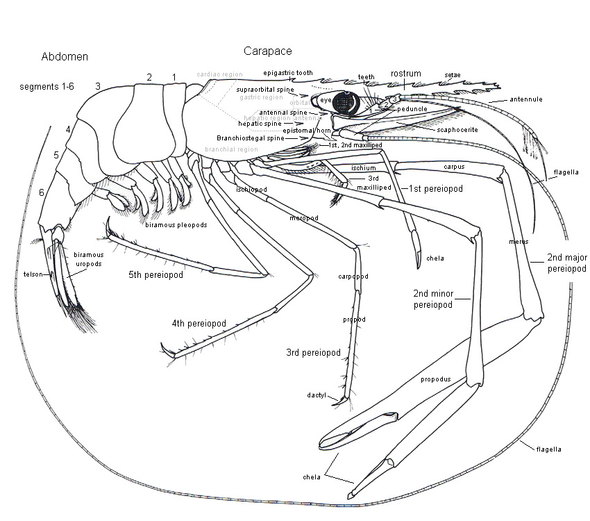
Figure 1
Glossary of common terms used in taxonomy:
cf : (example - Genus name cf species name) - denotes a case where the identity is in doubt, but the organism is similar to a known species in the opinion of whoever "cf-ed" them. Sometimes these end up being assigned to the names species, sometimes they end up being new species. cf = confer (Latin), = compare (English).
nov: New as in a new species.
aff. (affine): with affinity to.
species complex: A group of species which satisfy the biological definition of species, that is, they are reproductively isolated from each other, but they are not morphologically distinguishable or at least are not readily or reliably distinguishable on a morphological basis.
Glossary of descriptive terms with notes on form and function :
References:
Bauer, Raymond T. Remarkable shrimps : adaptations and natural history of the Carideans. University of Oklahoma Press, Norman, Publishing 2004

cf : (example - Genus name cf species name) - denotes a case where the identity is in doubt, but the organism is similar to a known species in the opinion of whoever "cf-ed" them. Sometimes these end up being assigned to the names species, sometimes they end up being new species. cf = confer (Latin), = compare (English).
nov: New as in a new species.
aff. (affine): with affinity to.
species complex: A group of species which satisfy the biological definition of species, that is, they are reproductively isolated from each other, but they are not morphologically distinguishable or at least are not readily or reliably distinguishable on a morphological basis.
Glossary of descriptive terms with notes on form and function :
Abdomen: (Fig.1) The tail, consisting of six body segments and the telson/uropods.
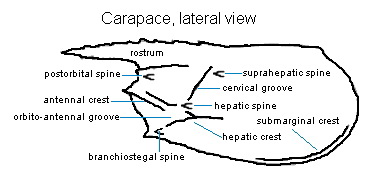
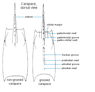
Adrostral Carina: The ridge flanking the rostrum, sometimes nearly reaching the end of the carapace.
Adrostral Sulcus: The groove flanking the rostrum to the adrostral carina, sometimes nearly reaching the end of the carapace.
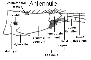
Antenna (Antennae): (Fig.1) Long, paired, usually flagellate appendage projecting from the front of the cephalothorax.
Antennal Carina: The ridge extending posteriorly along dorsal extremity of antennal region, often continuous with antennal spine.
Antennal Flagellum (Antennal Flagella): (Fig.1) Multiarticulate, whip-like terminal part of the antenna.
Antennal Peduncle: Three basal segments of the antenna, from which the flagellum arises.
Antennal Region: Area on the lateral face of the carapace posterior to and encompassing the antennal spine.
Antennal Spine: Spine situated on the anterior margin of the carapace just ventral to the orbital margin.
Antennular Flagellum (Anmnnular Flagella). Multiarticulate paired filaments (sometimes flattened and lamellate) of the antennule.
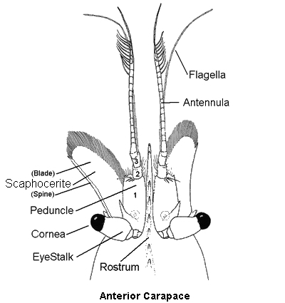
Antennular Peduncle: Three basal segments of the antennule, from which the flagella arise.
Antennule: Short, paired, usually flagellate appendages projecting from the front end of the cephalothorax.
Anterior: The front or frontal area.
Anterolateral Carina: Longitudinal ridge extending along the anterior part of the carapace, ventral to the gastro-orbital carina.
Arthrobranchia (Arthrobranchiae): Branchia (gill) attached to the joint area between the body and the first podomere of the leg.
Article: Any one of the subdivisions of an appendage segment.
Articular Membrane: Uncalcified integument at a joint, permitting movement of the exoskeleton, as between the segments of a pereopod.
Basial Spine: Spine projecting from a thoracic appendage.
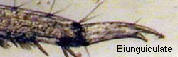
Biunguiculate: Having two of or being forked.
Branchia (Branchiae): Respiratory organ (gill) associated with an appendage or with the body wall.
Branchial Region: Area of the carapace overlying the branchial cavity and gills.
Branchiocardiac Carina. Ridge extending along the posterodorsal limit of the branchiostegite.
Branchiocardiac Sulcus: Groove extending along the dorsal limit of the branchiostegite, running parallel to the branchiocardiac carina.
Branchiostegal Carina: Longitudinal ridge extending along the anteroventral part of the carapace, usually continuous with the branchiostegal spine.
Branchiostegal-Hepatic Carina: Longitudinal ridge consisting of the fusion of the branchiostegal and hepatic carinae.
Branchiostegal Spine: Short spine on or near the anterior margin of the carapace ventral to the antennal spine and dorsal to the anteroventral angle of the carapace.
Branchiostegal Sulcus: Groove often accompanying the branchiostegal carina, located on the anteroventral part of the carapace.
Branchiostegite: Expanded ventrolateral part of the carapace covering the gills.
Carapace: The "head shield" cuticular structure arising from the posterior margin of the cephalon, extending anteriorly and posteriorly, and covering the cephalothoracic somites of the body.
Carina (Carinae): A ridge or keel of the exoskeleton.
Cephalothorax: Anterior part of the body consisting of the fused cephalon (head) and thorax, bearing all the appendages except the pleopods and uropods.
Cervical Carina: Transverse ridge starting at the top of the carapace and extending down and toward the front of the carapace.
Cervical Sulcus: Transverse groove starting at the top of the carapace and extending down and toward the front of the carapace.
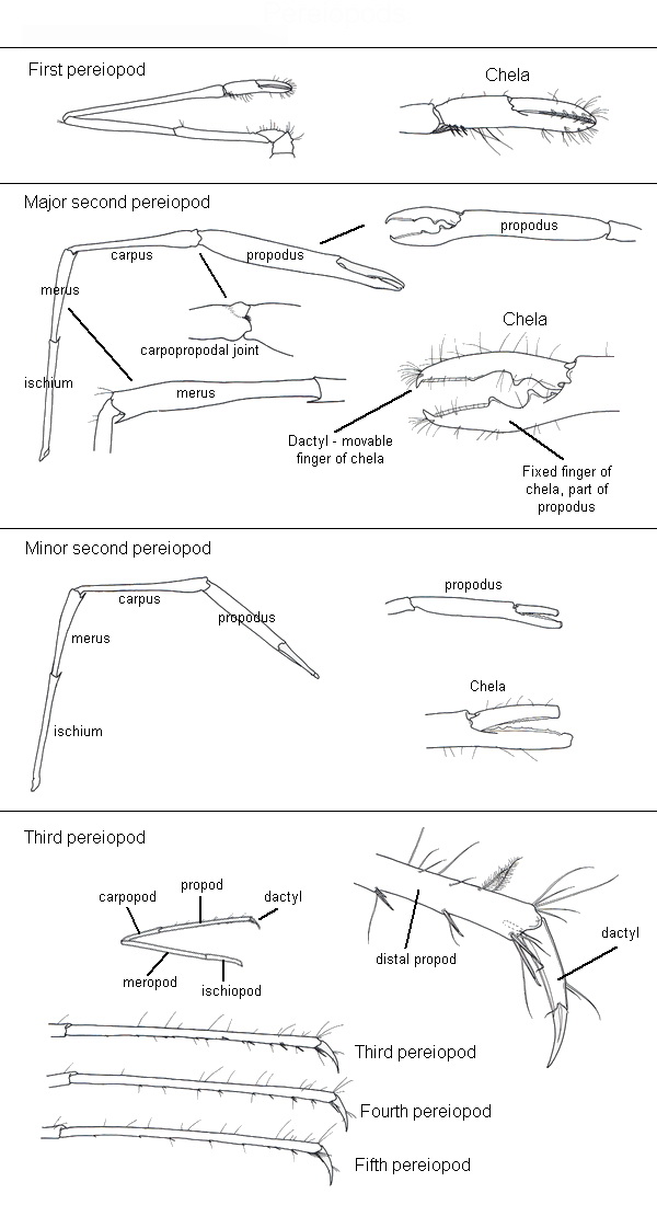
Chela (Chelae): Pincer formed by the two distal podomeres of a pereopod in which the movable finger (dactyl) opposes a fixed finger formed by a distal extension of the propod.
Chelate: Appendage ending in a chela (claw).
Cheliped: Any chela (claw)-bearing thoracopod; typically refers to first pair(s) of pereopods. (See below Photos)
Cicatrix (Cicatrices): Longitudinally disposed ridge often present on the lateral part of the sixth abdominal somite.
Cincinnuli (Cincinnulus): Minute interlocking processes projecting from the dorsomesial margins of the petasmal endopods.
Cornea: Faceted, usually pigmented portion of the eye.
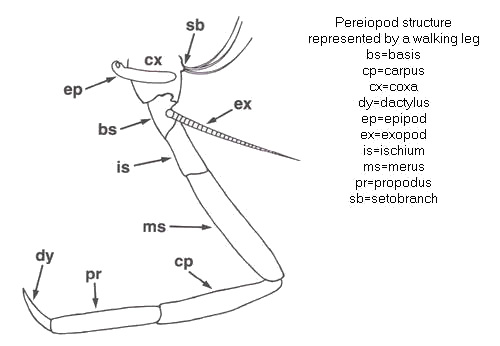
Coxa (Coxae): First or proximal podomere of a typically seven-segmented appendage.
Coxal Spine: Spine projecting from the coxa of a thoracic appendage.
Dactyl: Terminal podomere of a typically seven-segmented appendage.
Dendrobranchiate Gill: One in which the paired primary branches are subdivided, sometimes highly so.
Discoidal: Cleavage or furrow.
Distal Fold: Distal pleat in the dorsolateral lobule of the petasma.
Distolateral Projection: Distolateral, ventrally inclined projection or spur of the basis of the endopod of the male second pleopod.
Distomedian Projection: Distal, relatively narrow extension of the dorsomedian lobule of the petasma.
Dorsolateral Carina: Longitudinal ridge on the dorsolateral region of the carapace running dorsal to the orbital region.
Dorsolateral Lobule: Dorsal part of the lateral lobe of the petasma.
Dorsolateral Sulcus: Longitudinal groove sometimes present close to the dorsomedian line of the sixth abdominal somite.
Dorsomedian Carina: Ridge extending along the middorsal line of the abdominal somites.
Dorsomedian Lobule: Part of the median lobe of the petasma.
Endopod: Mesial ramus of a biramous appendage, especially one arising from the basis or from the protopodite of the pleopod.
Endite: Lobe of several proximal podomeres of various appendages.
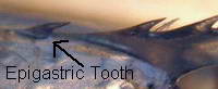
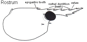
Epigastric Tooth: Tooth on the carapace situated above the gastric region behind the first (posteriormost) rostral tooth.
Epipod: Lateral exite of the coxa of a thoracic appendage, sometimes branchial in function.
Epistome: Transverse plate anterior to mouth area.
Exopod: Lateral ramus of a biramous appendage, arising from the basis, or from the protopodite.
Exuviae: The shed exoskeleton or molt.
Eyestalk: Peduncle or unfaceted part of the eye supporting the cornea.
Flagellum (Flagella): Multiarticulate, usually whip-like terminal part of the antennule or antenna.
Frontal Region: Anterior area of the carapace lying between the orbits and bounded posteriorly by the gastric region.
Gastric Region: Principal median area of the carapace bounded anteriorly by the frontal and orbital regions, and posteriorly by the cardiac region, and laterally by the branchial and hepatic regions.
Gastrofrontal Carina: Short longitudinal ridge extending posteriorly from the ventral extremity of the orbital region.
Gastrofrontal Sulcus: Short longitudinal depression accompanying the gastrofrontal carina dorsally.
Gastrohepatic Gland: Digestive gland associated with the midgut, within the cephalothorax.
Gastro-Orbital Carina: Short longitudinal ridge extending (often curving) anterodorsally from the cervical sulcus towards the orbital region.
Genitalia: The external reproductive structures.
Glabrous: Smooth, glossy.
Hepatic Carina: Longitudinally or obliquely disposed ridge of variable length lying ventral to the hepatic region, sometimes extending almost to the anterior margin of the carapace.
Hepatic Region: Paired anterolateral areas of the carapace bounded anteriorly by the antennal region, posteriorly by the branchial region, and mesially by the gastric region.
Hepatic Spine: Lateral spine situated near the anterior margin of the hepatic region of the carapace.
Hepatic Sulcus: Groove ventral to the hepatic region extending posteriorly, sometimes from near the anterior margin of the carapace.
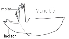
Incisor Process: Cutting process, often toothed or cusped, of the mandible.
Insemination: The act of placing or introducing sperm onto or into the thelycum or seminal receptacles of the female
.
Integument: Outer covering or exoskeleton.
Ischial Spine: Spine projecting from the ischium or third segment of the thoracic appendage.
Ischium (Ischia): Third podomere from the proximal end of a typically seven-segmented appendage.
Labrum: Upper lip or unpaired structure arising anterior to the mouth and often covering it.
Lateral Lobe: One of the paired lateral parts, often folded, of the petasma.
Longitudinal Suture: Fine longitudinal line extending posteriorly just above the base of the antennal spine.
Mandible: One of the heavily calcified jaws lying anterior to (beneath, in ventral view) other mouth-parts. Each mandible is a stout, muscle filled structure and comes in several variations. It may carry both an incisor process for biting and cutting and a molar process for crushing and grinding food. Often present is a small, segmented palp (mandibular palp) equipped with setae which may function in cleaning the mouthparts. The various combinations of palp, incisor process and molar process are important features in the taxonomy and classification of the caridean shrimp.
Mandibular Palp: Segmented endopods attached laterally to the masticatory part of the mandible.
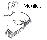
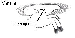
Maxillae: Posterior to the mandibles are two pairs of maxillae which play a role in food handling.
1st maxilla (maxillule) : The second mouthpart and fifth cephalic appendage.
2nd maxilla (maxilla): Paired mouthpart appendages of the fourth and fifth cephalic somites. The lateral blade of the maxilla (scaphognathite) extends back into the gill chamber, also known as the gill bailer.
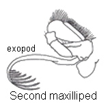
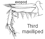
Maxillipeds: One of a pair of three sets of thoracic appendages, arising posterior to the primary mouthparts. The two anterior pairs are often modified for feeding, while the third pair is often pediform, resembling the pereopods.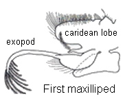
Median Lobe: One of the paired dorsal parts, often folded, of the petasma.
Median Sulcus: Dorsomedian groove on the carapace.
Merus (Meri): Fourth segment from the proximal end of a typically seven-segmented appendage.
Mesial Tubercle: A conical to low, rounded protuberance on the surface of the optic calathus.
Ocular Plate: Median cephalic plate bearing the eyestalks laterally.
Ocular Scale: Scale-like structure located on the basal segment of eyestalk.
Optic Calathus: Terminal article of the eyestalk supporting, often embracing, the cornea of the eye.
Orbital Margin: Anterior border of the carapace, often contiguous to the eye.
Orbital Region: Paired areas on the carapace just posterior to the eyes.
Orbital Spine: Spine projecting from the ventral extremity of the orbital margin.
Orbito-Antennal Sulcus: Longitudinal or oblique depression between the orbital margin and the hepatic spine.
Palm: Portion of the chela proximal to the propodal finger.
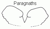
Paragnaths: A pair of ventral projections of the cephalic cuticle just posterior and medial to the mandibles.
Pereiopod: One of the five posterior paired appendages or legs of the cephalothorax. These limbs have the typical coxal and basal segments of a biramous crustacean limb and a sticklike endopod of five segments: ischium, merus, carpus, propodus and dactylus (dactyl). The first two pairs of caridean pereopods are equiped with chelae (claws). The chelae are used for food searching and handling, aggression and defense and grooming. In many caridean species one of the two pairs is stout and robust while the other pair is slender and delicate with the chelae carrying tufts of setae which can be used for grooming. The posterior (or last) three pairs of pereopods are the walking legs, allowing the shrimp to step in all directions as well as supporting the body when at rest. There is considerable variation in the length and thickness of the walking legs amongst the species and usually correlate with the general body robustness and exoskeleton thickness.
Petasma (Petasmata): The male genital structure consisting of the much enlarged and coupled endopods of the first pair of pleopods.
Petasma, Open: The lateral lobes are quite flexible, partially or entirely extended laterally, with the ventral costae not or barely turned ventrally.
Petasma, Semi-Open: The lateral lobes are flexible but folded, with the ventral costae distinctly turned ventromesially, delimiting relatively ample space extending from proximal to distal ends.
Petasma, Semi-Closed: The lateral lobes are rather flexible, markedly folded, supported by strong ribs, with the ventral costae approaching rather closely, delimiting a moderately large space, narrowly open distally where usually overlapped by well-developed distomedian projections.
Petasma, Closed: The lateral lobes are heavily sclerotized, sometimes making the structure virtually rigid, with the ventral costae situated ventromesially, almost abutting, and delimiting a small, sometimes extremely so, space; lateral lobe is usually produced distally into lateral spouts or horns.
Phyllobranchiate Gill: A gill in which the branches are plate-like, usually occurring in paired series along the gill axis.
Pleopod: One of the biramous paired appendages typically arising ventrally from each of the anterior five abdominal segments. They are primarily swimming organs.
Pleurobranchia (Pleurobranchiae): Gill attached to the body wall (pleural membrane), dorsal to the articulation of the appendage.
Pleurite : One of the lateral flaps on each of the anterior five abdominal segments.
Podobranchia (Podobranchiae): Gill borne on the basal segment (coxa) of a thoracic appendage.
Podomere: Any one of the segments of an appendage, such as a segment of a pereopod or maxilliped.
Postantennal Spine: Spine located on the anterolateral area of the carapace (on the posterior part of the antennal region).
Postcervical Spine: Spine located immediately posterior to the cervical carina.
Postcervical Sulcus: Subvertical carapace groove located posterior to the cervical sulcus.
Posterior: The polar opposite of Anterior (front) or the back end.
Posterior Process: Posterior part of an elongate median protuberance projecting caudally onto the last (XIV) thoracic sternite.
Posterior Protuberance: Conspicuous elevation arising from the posteromedian part of the last (XIV) thoracic sternite.
Posthepatic Carina: Ridge posterior to the hepatic carina, extending onto the lower branchiostegite.
Postocular Sulcus: A small groove situated near the dorsal extremity of the orbital margin.
Postorbital Spine: Spine situated near the orbital margin posterior to the antennal spine.
Postrostral Carina: Dorsomedian ridge extending posteriorly from the base of the rostrum, sometimes nearly reaching the posterior margin of the carapace.
Propodus (Propodi): Sixth or penultimate segment of a typically seven-segmented appendage.
Prosartema: Variable in shape, thin, sometimes scale-like process arising from the mesial base of the first antennular segment, and extending distally.
Protocephalon: Anteriormost part of the body bearing eyes.
Pterygostomian Carina: Ridge running posterior to pterygostomian spine on anteroventral part of carapace.
Pterygostomian Region: Anteroventral area of the carapace.
Pterygostomian Spine: Marginal spine arising from the anteroventral angle or border of the carapace.
Ramus (Rami): A branch of an exopod or endopod.
Rostrum (Rostra): Anteromedian projection of the carapace between the eyes. (See below Photos)
Scaphocerite: Laterally rigid lamellate exopod of the antenna; the antennal scale.
Segment: Division of an appendage.
Seminal Receptacles: Unpaired or paired bulbous or tubular sacs associated with the thelycum for the storage of spermatophores or sperm.
Unpaired Receptacles: Noninvaginated, opening either through an exposed median longitudinal slit flanked by lateral plates of sternite XIV, or opening anterior to a single plate.
Paired Receptacles: Invaginated into the cephalothoracic cavity, opening through well protected slits usually on anterior border of sternite XIV, or on anterior part of XIII.
Somite: Each of the main divisions of the body.
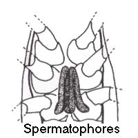
Spermatophore: The sperm-carrying, variously complex mass, issuing from the male petasma during copulation.
Statocyst: Sensory organ of awareness of rotation and position located at the base of the first antenna.
Sternite: Ventral part of a thoracic or abdominal somite.
Sternum: Ventral surface of the cephalothorax or abdomen.
Stylocerite: Pointed scale arising from the lateral base of the first segment of the antennular peduncle.
Subhepatic Sulcus: Groove located well ventral to the hepatic region of the carapace and the hepatic spine.
Sublateral Sulcus: Groove located ventral to the lateral carina of the carapace.
Submarginal Carina: An almost longitudinal ridge extending between the rigid and the membranous part of the branchiostegite.
Subrostral Sulcus: A longitudinal elongate groove extending along the dorsal limit of the orbital region.
Sulcus (Sulci): Groove.
Suprahepatic Spine: Spine arising from the edge of the cervical carina dorsal to the hepatic spine.
Supraorbital Spine: Spine located posterior to the orbital margin of the carapace.
Suture: Either transverse or longitudinal, weakly sclerotized line or seam on the carapace.
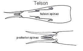
Telson: Terminal unit of the abdomen bearing the anus.
Tergum (Terga): Arched dorsal part of each of the anterior five abdominal somites.
Thelycum (Thelyca): The female genitalia consisting of modifications of the posterior two, or sometimes three, thoracic sternites (XII–XIV) serving for the storage or transfer of the sperm, usually in spermatophores, and often shielding seminal receptacles.
Thelycum, Open: One in which the seminal receptacles are absent.
Thelycum, Closed: One in which the seminal receptacles are present.
Thoracic Ridge: Highly sclerotized, rib-like transverse structure across posterior margin of sternite XIV.
Transverse Suture: Fine, short vertical line extending dorsally from the ventral margin of the carapace.
Trichobranchiate Gill: A gill in which the branches are fingerlike and project from a central axis.
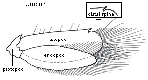
Uropod: Paired, biramous appendage attached to the sixth abdominal segment, usually combining with the telson to form a tailfan.
Ventral Costa: Ridge extending along the ventropiesial margin of the ventrolateral lobule of the petasma.
Ventral Surface: The side closest to the ground or the "under-side".
Ventrolateral Lobule: Ventral part of the lateral lobe of the petasma.
Ventromedian Lobule: Lateral part of the median lobe of the petasma.

References:
Bauer, Raymond T. Remarkable shrimps : adaptations and natural history of the Carideans. University of Oklahoma Press, Norman, Publishing 2004